A new study led by Dr. Kai Wang from the Department of Ophthalmology, Peking University People's Hospital and Dr. Hongxin Song from Beijing Tongren Hospital has been published in JAMA Ophthalmology. This research focused on the effects of repeated low-level red light (RLRL) therapy on myopic children's retinas.
Background And Method
The global prevalence of myopia is on the rise, especially among children and adolescents. RLRL therapy has shown potential in controlling myopia progression, but its long-term impact on retinal photoreceptors remains unclear. To address this, the research team conducted a retrospective multicenter cohort study. They recruited 99 myopic children, divided into an RLRL group (52 children) and a control group (47 children). The team used the AOSLO system (Robotrak Technologies, Nanjing, China) to measure cone photoreceptor density along 4 retinal meridians. This system has a navigation system to help localize the imaging area and uses a multiplane independent refractor lens to correct wavefront aberrations, ensuring high-quality image acquisition.
Results
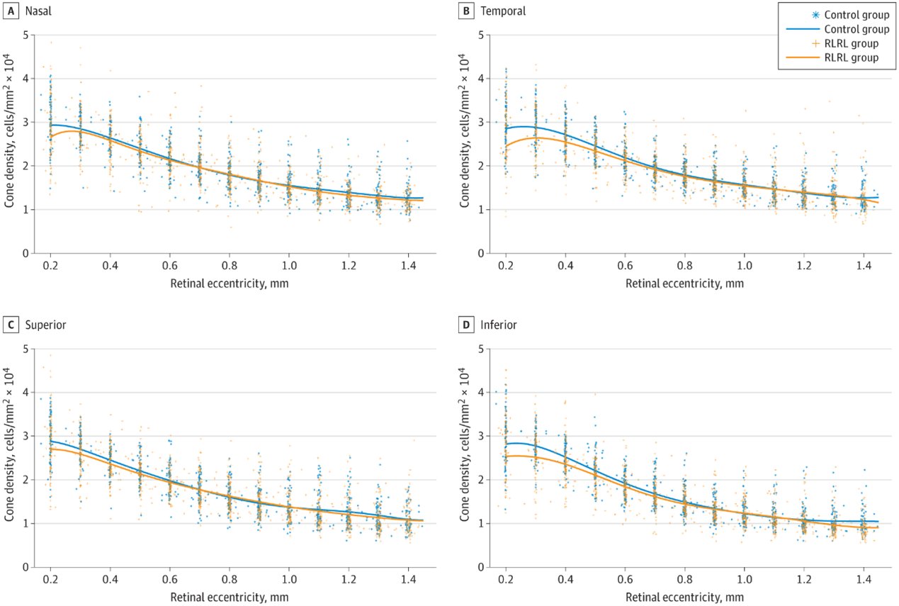
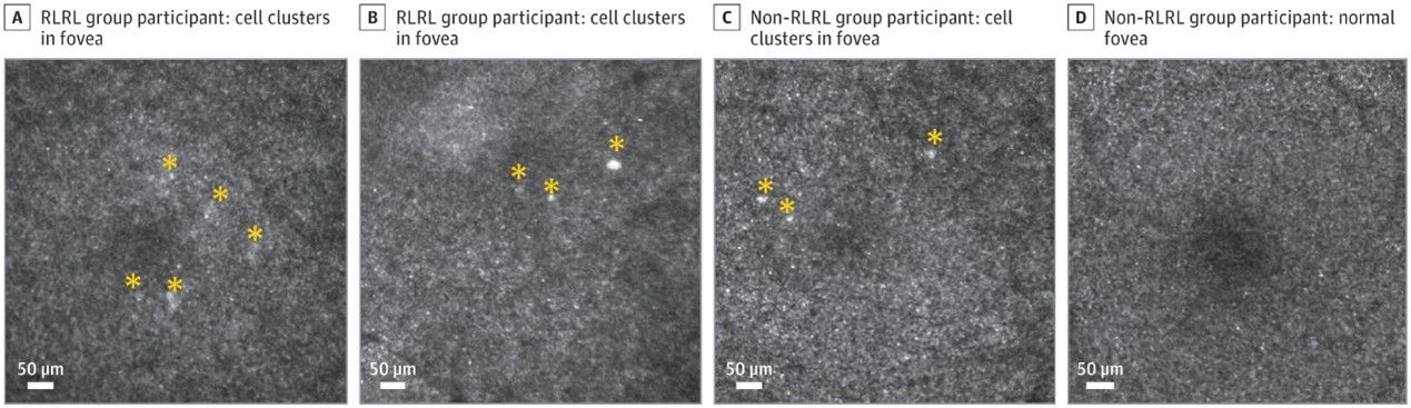
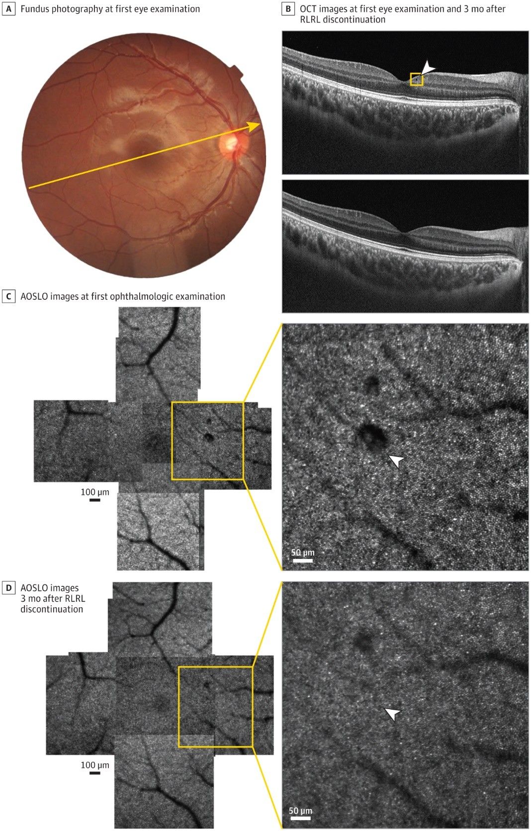
The study results showed that RLRL users had a decreased cone density within 0.5 mm eccentricity from the foveal center, especially in the temporal region. For example, at 0.3 mm temporal eccentricity, the RLRL group had a significantly lower cone density, with a mean difference of -2.1×10³ cells/mm² compared to the control group (P = 0.003). Additionally, 11 eyes of 10 participants showed abnormal low-frequency, high-brightness signals near the fovea, and the odds ratio of such abnormal signals in RLRL users was 7.23 times higher than non-users (P = 0.02). One participant in the RLRL group had small cystoid abnormalities in the ganglion cell layer, which disappeared 3 months after discontinuing RLRL therapy.
Summary
This study indicates that RLRL therapy for at least 1 year may be linked to reduced cone density in the paracentral fovea and other subtle retinal abnormalities in some myopic children. Although RLRL therapy shows promise in controlling myopia, its long-term safety and efficacy need further exploration. AOSLO assessment may serve as an important tool for evaluating the efficacy and safety of RLRL therapy.